Cambridge

Medical Tripos 1A

The Basics

The 3 years of your undergraduate at Cambridge can be divided up into parts 1A (first year), 1B (second year), and part II.

1A encompasses 3 major subjects, Functional Architecture of the Body (FAB), a.k.a Anatomy, Molecules in Medical Science (MIMS), aka Biochemistry, and Homeostasis (HOM), which is physiology.

FAB

  • You’ll have 2 dissections in one week, and then 1 dissection the following week
  • There will be lectures that run in parallel, these can also be tested in MCQs
  • I’d recommend memorising the parts of the manual corresponding to each session before the sessions -> this really helps you to get the most out of dissection
  • Course Breakdown
    • Upper Limb – really make sure you know the origins and attachments of the muscles, this is very valuable for the MCQs
    • Thorax – probably the easiest topic there is, very easy to visualise
    • Abdomen and Pelvis – the hardest topic to visualise, it’s worth putting in the work to make sure you understand this during term, because it’ll be a pain to go over it in the holidays
    • Lower Limb – same advice as for upper limb, this topic is probably the smallest one
    • Embryology – these lectures run in parallel with what is covered during dissection, and also can come out in the MCQs

MIMS

  • 3 lectures a week, 1 practical a term (with a discussion session 2 weeks later), 2 PBL presentations across Michaelmas and Lent 
  • Michaelmas
    • You’ll start off with an introduction to diabetes, with 5 lectures on protein structure and enzyme activity
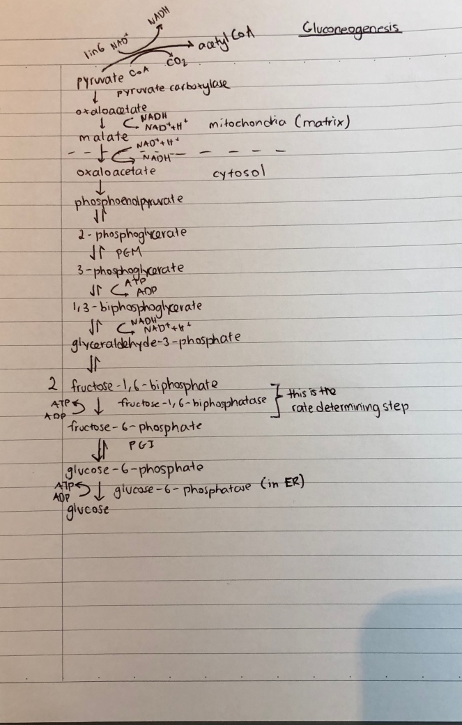
    • After this, the more nitty gritty stuff comes in, in the form of metabolism
    • This carries on for a while, the term finally ends off with cell signalling and protein sorting, the former is very useful for HOM, so I’d make an effort to try to learn this during term
  • Lent
    • The major theme of this term is molecular biology and the clinical linker is cancer, so the prologue and epilogue lectures will be centered on this
    • You’ll start off with the basics of the genome, then gene expression, followed by broad genetics 
    • The term ends off with the cell cycle and cell death 
    • There are also 2 lectures just on cancer; you can build on these for essays to score.
  • There is a lot of very specific detail you are expected to know, so build a system over the Christmas vacation that lets you learn it efficiently 
  • The practical paper is quite challenging, and doesn’t really get much attention in the practical sessions themselves, so I would recommend practising these over the Easter vacation
  • Make a list of experimental techniques so when you’re asked to suggest further experiments, you can jot down some good ideas when asked in the practical paper

HOM

  • 3 lectures a week
  • HOM doesn’t require as much brute force memorisation as MIMS or FAB, but you do have to make an effort to wrap your head around the logic of certain concepts. 
  • These are the major topics with some advice for each
  • Nerves
    • Quite a mathematical topic, you can usually relate most concepts to the Nernst equation, which I’d recommend understanding the derivation of
    • Learn the experiments; they could test them in MCQs, or you could use them in essays
    • Supervisions should give you some additional diagrams to use in essays, the ones brought up in lectures are a bit basic 
  • Muscles
    • Create a table comparing the different kinds of muscles, it helps to produce a nice mental schema for compare and contrast essay questions
  • Cardiovascular
    • Very content heavy topic, but well taught 
    • Make sure you understand Starling’s Law, because a good understanding of it helps for both respiratory and renal 
  • Respiratory
    • Get your head around what increases/decreases compliance early on, it makes life much easier later 
    • There are a lot of numbers to know for this topic, so make sure you use the right units for them
  • Renal
    • Again, very content heavy, and it’s the longest lecture series
    • Make a table for all the transporters, and for hyper and hypokalaemia/calcaemia
    • Get the Koeppen monograph – it explains stuff clearly and has loads of extra stuff you can use in essays 
  • Digestion
    • One of the easiest HOM topics, but the most content per unit lecture
    • Create a table for the GI hormones
  • Endocrine
    • This is actually a pretty good lecture series, probably worth going for, even though it’s in Easter

Some of My Resources

FAB

  • A set of tables I made summarising the anatomy of the upper limb, with blanked out tables by the side for testing

MIMS

HOM

Have fun!

Biology

Gluconeogenesis

One of the pillars of cellular metabolism is that, during anaerobic respiration, lactate is produced at the end of glycolysis following the reduction of pyruvate by lactate dehydrogenase A. It is the reason we have an “oxygen debt” to fulfil following periods of intense exercise. However, is oxidising lactate the only way we can remove lactate from the blood? Can we make more functional use of it?

The answer to the latter is, surprisingly, yes. To set the stage, let’s consider the enzymes involved in glycolysis. Interestingly, all of them, with the exception of hexokinase and phosphofructokinase, can catalyse the reverse reactions of glycolysis.

Lactate from the muscles is released into the blood, and subsequently is taken up by the liver, which has the necessary enzymes for a process called gluconeogenesis. Etymologically, “gluco” refers to glucose and “neogenesis” refers to the regeneration of a substance.

During gluconeogenesis, lactate is converted back into pyruvate and subsequently, this pyruvate is converted into oxaloacetate in the mitochondria. The oxaloacetate is then reduced to malate, which is transported out of the mitochondria, into the cytoplasm, where it is oxidised to reform oxaloacetate. The oxaloacetate is then decarboxylated and phosphorylated to form phosphoenolpyruvate.

This is where things get very unusual – the phosphoenolpyruvate undergoes what can be best labelled as “reverse glycolysis,” as illustrated below.

IMG_1372.jpg

It reforms glucose, which is released into the blood and taken up by actively respiring muscle fibres to produce the ATP needed for the sliding of the actin filaments in those muscle fibres (specifically to enable the cocking of the myosin heads and the breaking of the crossbridges between the myosin heads and the actin binding sites). This process of recycling lactate to reform glucose is called the Cori Cycle.

As I’ve mentioned on the diagram, one in every 6 pyruvates goes through the link reaction to produce acetyl CoA, which helps to produce the reduced hydrogen carriers needed for oxidative phosphorylation. This produces the needed ATP to fuel gluconeogenesis and the Cori Cycle.

Interestingly, gluconeogenesis is not a process limited to liver cells; renal cells (most prominently in superficial cortical nephrons) as well as astrocytes in the brain can carry out gluconeogenesis, likely to enable them to meet their atypically high ATP requirements.

I’ve attached a document here, with a (admittedly) more verbose explanation of how gluconeogenesis occurs, with some forays into other aspects of glucose metabolism as well.


Biology

Phototransduction

An interesting concept I’ve stumbled across during my reading into neurobiology is that a neurotransmitter need not be exclusively excitatory or inhibitory. In fact, a prime example of this would be glutamate in phototransduction, the process by which the stimulation of photoreceptors by photons triggers the propagation of an action potential across the optic nerve.

retina diagram

A pretty good minimalist diagram from Bioninja that shows the basic structure of the retina


The behaviour of photoreceptors in the dark

In the dark, we find that the photoreceptor cells have a relatively high membrane potential (-40mV). This membrane potential comes about because, in the dark, these cells have high concentrations of a molecule called cyclic guanosine monophosphate (cGMP), which binds to cGMP gated Na+ and Ca2+ channel proteins to allow their entry by facilitated diffusion into the photoreceptor, hence the cell to exhibit a depolarised membrane potential.

This has the effect of keeping voltage gated Ca2+ channels between the synaptic knob and the main body of the photoreceptors open, thus facilitating the movement of glutamate across the synaptic cleft and allowing it to bind to receptors on the post-synaptic bipolar cell.

This is where things get interesting. Glutamate is typically a fast-acting, excitatory neurotransmitter, but in this particular case, it behaves as a slow-acting inhibitory neurotransmitter. I initially thought it was able to do this by binding to a glutamate gated anion channel.

As it would seem, glutamate activates a cytoplasmic guanosine (G) protein by binding to receptors on the membrane of the bipolar neuron. This G proteins initiates a cascade of reactions that reduce the cytoplasmic cGMP concentration, thus reducing binding of cGMP to cGMP gated cation channels that need to be open to trigger depolarisation.

As a result, in the dark, the bipolar neuron is hyperpolarised (its membrane potential is approximately -65mV) due to the glutamate released by the depolarised photoreceptors. The bipolar cell’s inability to depolarise is what inhibits any impulses from being transmitted from the eye to the brain under pitch-black conditions.

What is fascinating is how the membrane potential across the bipolar cell changes when light strikes the photoreceptor beneath it.


The behaviour of photoreceptors under illumination

In regions called receptor disks, photopigments are stored within photoreceptor cells. In rods, the main photopigment is rhodopsin – a molecule associated with retinal and a number of opsin polypeptides. When retinal absorbs a photon, its configuration changes from its 11-cis isomer to an all trans isomer. This initiates a series of reactions which result in the activation of the intracellular signalling protein transducin (from which phototransduction derives its name).

Transducin in turns activates phosphodiesterases which hydrolyse cGMP molecules. This has the effect of reducing the concentration of cGMP in the photoreceptors, thus reducing binding to cGMP gated Na+ and Ca2+ channel proteins, thus resulting in hyperpolarisation of the photoreceptor cell.

This decrease in membrane potential closes the voltage gated Ca2+ channels, reducing the concentration of Ca2+ in the synaptic knob. As a result, the quantity of glutamate released sharply decreases, leading to the depolarisation of the bipolar cell, triggering the propagation of an action potential along the axons of the ganglia.

Moving away from neurotransmitters momentarily, let’s look into how cytoplasmic Ca2+ concentrations can influence the extent to which photoreceptors respond to light.


Sensitivity to Light Intensity

Generally, photoreceptors display diminishing sensitivity to illumination as light intensity increases. When the Ca2+ concentration in the photoreceptor decreases, it triggers a small change in the phototransduction cascade, by initiating the upregulation of the enzyme that produces cGMP and also increasing the affinity the cGMP gated Na+ and Ca2+ channels have for cGMP. This reduces the sensitivity of the photoreceptors to any increases in illumination by requiring a larger number of activated photopigments to reduce the cytoplasmic cGMP concentration to a low enough level such that the membrane potential can be decreased.


Recycling of retinal

With respect to the photopigments, there must always be a sufficient quantity of viable photopigments in the receptor disk. To ensure that this is the case, following the depolarisation of the photoreceptor, the protein arrestin binds to activated rhodopsin molecules, preventing them from binding to transducin and facilitating their movement to the pigment epithelium.

In the pigment epithelium, a series of enzymatic reactions convert the all trans retinal molecule in rhodopsin into its 11-cis form. Following this, the rhodopsin molecule moves back into the receptor disk. This system effectively allows for the recycling of previously activated photopigments and maintains the presence of inactivated photopigments in the receptor disk.


Conclusion

Phototransduction is an example of a process whose beauty lies in the rationality of its design – it continuously regenerates new components, gives rise to variable light sensitivity, and most importantly, allows us to see.

Biology

Hypoxia Inducible Factors

It is axiomatic that a lack of oxygen often proves fatal to animals, but it is interesting to note how animals can adapt their biochemistry to suit this environment.

The mechanism by which this occurs in most animals is fascinating – it relies on a family of transcription factors named Hypoxia Inducible Factors (HIFs). HIFs are typically inactive in cells with adequate oxygen concentrations. This is because hydroxylases, enzymes that add hydroxyl groups to HIFs, have oxygen available to them as co-substrates, hence an O-H bond can be formed and the HIFs can be hydroxylated.

The hydroxyl group acts as a signal for recognition proteins, thereby targeting the HIFs for degradation. However, in low oxygen concentrations, the co-substrate concentration levels decrease, leading to fewer products being formed per unit time, hence leaving a larger number of HIFs intact.

When stabilised by the lack of oxygen, the HIFs bind upstream of the promoters of various genes. For example, it upregulates the transcription of hexokinase and phosphoglucose isomerase, 2 enzymes involved in glycolysis. This facilitates the production of ATP in an anaerobic environment, hence maintaining a supply of ATP for the cell.

In addition, HIFs upregulate the transcription of the vascular endothelial growth factor (VEGF), which enables angiogenesis – the formation of new blood vessels. This has the effect of allowing greater blood flow to actively respiring tissues in low oxygen conditions, such as muscle cells.

The role of HIFs in triggering angiogenesis has not gone unnnoticed by cancer scientists. Research which looks promising is currently being carried out into the effectiveness of HIV inhibitors in the context of metastatic cancers (http://onlinelibrary.wiley.com/doi/10.1111/j.1582-4934.2009.00876.x/full).

Biology

RAAS (Renin Angiotensin Aldosterone System)

Having done a bit of reading into hypertension recently, I stumbled upon this homeostatic pathway quite unintentionally, but found it fascinating.

RAAS starts off with renin, which is a proteolytic (protein “breaking”) enzyme produced by juxtaglomerular cells in the afferent arteriole of the kidney. Renin is mainly produced by these cells as a homeostatic response to hypotension. The sympathetic autonomic system detects lower systemic vascular resistance and signals beta adrenoceptors in the juxtaglomerular cells to produce renin. The produced renin is subsequently circulated around both the systemic and pulmonary circulations.

IMG_9621.jpg

A rather poorly drawn diagram depicting the RAAS system (the core processes)

Renin acts on a circulating substrate – angiotensinogen, which is broken down into the decapeptide (10 amino acids) angiotensin 1. This is where ACE (Angiotensin Converting Enzyme) comes into play. ACE is produced in the vascular endothelium, particularly within the blood vessels in the lungs. It acts on angiotensin I, producing angiotensin II by removing isoleucine and valine.

Now things start to get interesting.

Angiotensin II has a myriad of functions, the most notable of which are as follows.

  1. Stimulates resistance vessels to constrict, increasing systemic vascular resistance and thus raising blood pressure
  2. Triggers Na+ reabsorption in the distil convoluted tubule directly
  3. Initiates aldosterone secretion by the adrenal cortex, thus indirectly triggering Na+ reabsorption in the distil convoluted tubule
  4. Stimulates the posterior pituitary gland to release vasopressin (ADH), to increase water reabsorption
  5. Stimulates the thirst centres of the brain
  6. Triggers cardiac and vascular hypertrophy

Therefore, it makes sense that ACE inhibitors would also be useful anti-hypertension drugs, since they reduce the concentration of angiotensin II in the blood, therefore limiting increases in blood pressure. Additionally, ACE inhibitors also are used in nephrology to treat patients with diabetes induced chronic kidney disease (sometimes called diabetic nephropathy), since it reduces further damage to the glomeruli.

So, all you would-be-doctors reading this, I’d recommend keeping this pathway tucked away in your head if you want a leg up as a medical student.


Biology · Chemistry

What’s a good example of an interesting amino acid?

glycine.png

What a fascinatingly simple molecule. We typically assume that long, complex molecules with equally long names have the most interesting roles to play in Biochemistry, but this non-essential amino acid in itself offers the converse argument. So what makes glycine so special?

Firstly, its behaviour in acid-base reactions is two-fold; it can act as an acid or a base. In low pH solutions, it is protonated very easily, with a pKa of 2.4, to form CH­2COOHNH3+, whereas in high pH solutions, it forms CH­2NH2COO­, with a pKb of 4.4. This is one of the features that allows glycine to be featured prominently in a wide myriad of proteins (interestingly, collagen is 35% glycine) and polypeptide chains; depending on the pH of the surroundings, the same protein could take up different conformations, partially due to the amphoteric nature of glycine.

Now, let’s consider a more interesting role that glycine plays in mammalian systems. It acts as an inhibitory neurotransmitter – a substance that, when it binds to ionotropic post-synaptic receptors (transmembrane proteins that allow specific ions to enter a cell), results in the generation of an inhibitory post synaptic potential. What is interesting is the mechanism that initiates this. When glycine moves across the synaptic cleft and binds to ionotropic receptors on chlorine channels, said channels open, resulting in an influx of Cl ions into the post-synaptic neuron. This has the effect of reducing the cell potential far below the resting potential and even further below the threshold potential, creating an inhibitory post-synaptic potential. It is inhibitory because it reduces the likelihood of the threshold potential being released when a non-inhibitory neurotransmitter binds to the post-synaptic neuron, thus inhibiting the propagation of an action potential across a synapse.

Aside from playing a role in the regulation of the central nervous system, glycine is also broken down through a series of metabolic pathways to provide the C2N subunit for purines in DNA. 2 ways in which glycine is broken down are especially interesting, because of how they aid another key biological process in the human body – aerobic cellular respiration. When glycine reacts with tetrahydrofolate and NAD+ in its most common catabolic pathway, it ends up forming NADH. In another pathway, it is broken down to form serine, which is subsequently converted to pyruvate. Both NADH and pyruvate play key roles in aerobic respiration; the former is oxidised in the electron transport chain to facilitate oxidative phosphorylation and the latter is broken down in the link reaction to form acetyl coenzyme A, which is used in the Krebs’s cycle to produce NADH and FADH2.

Finally, it is the only amino acid that has been found in space, something that, arguably, could be traced to its simple structure.

Considering the breadth of roles glycine plays in mammals, its fascinating chemistry and presence in space; it certainly is an interesting amino acid.

Oncology

The Genetic Origins of Cancer

Personally, I am a strong proponent of the argument that cancer is inherently a genetic disease and I seek to explain why in this post.

Let’s start of with the basis of cell division. An important question here is, “why would a cell start dividing at a rate beyond the somatic level?” Well consider how every cell has checkpoints in place set up by specific genes to stop this happening. Of course there are other factors that could cause this, but in most cases it tends to be due to genetics. Consider the prevalence of TP53 mutations in cancers; these mutations tend to be present in almost all cancers.

So how could a mutation in this particular gene on chromosome 17 lead to rapid cell division? TP53 has a variety of functions, such as initiating G1 arrest if DNA damage is detected, triggering senescence if telomere lengths have become too low, starting a cascade that results in DNA repair proteins becoming active, inhibits angiogenesis and activates apoptosis. Hence, if this protein were absent or in a state in which it could no longer function, we would face a significant problem. Typically, mutations change the conformation of the p53 protein, leading to disfunction and possibly more detrimental effects, such as the promotion of sustained angiogenesis.

The significance of this disfunction is paramount – a change in the conformation of p53 could prevent it from binding to the promoter and promoter-proaxial elements of the gene coding for p21, a protein that extends the duration of time a cell spends in G1, hence granting the cell more divisions per unit time. This in turns leads to the development of more mutations, given than a mutation occurs every 10-10 bases per DNA replication. The change in the genetics of TP53 also has an effect on the ability of the cell to exceed the Hayflick limit (TERT also plays a role, but I will discuss this in another post), as cellular senescence may be delayed or even even disrupted completely if p53 does not function as it would in a somatic cell (Note that this assumes that the mutation also renders other proteins involved in cellular senescence useless). This demonstrates the significance of specific genes in the growth of cancers, which, by definition, start off as a mass of rapidly dividing cells.

To further illustrate my point, let me introduce another gene to the fold – RB1. RB1, otherwise known as the retinoblastoma gene, acts as a tumour suppressor – it can stop the progression of the cell cycle by preventing a cell from moving into the S phase. The protein it codes for, pRb (or p110), becomes active when DNA damage is detected and has a specific conformation that allows it to bind to the genes coding for E2F transcription factors that are active in DNA replication. pRb then attracts histone deacetylase to the site of the promoter, which loses its acetyl group due to the action of histone deacetylase. This is significant because it reduces the accessibility of the gene to transcription factors, thereby downregulating it and reducing the chance of the cell moving into the S-phase. However, if 2 mutated copies of the RB1 gene are present, pRb no longer functions as it would normally, possibly leading to cancer developing.

There is no dearth of other oncogenes I could refer to and explain but I believe my point has been made.